Online first
About the Journal
Current issue
Archive
Publication Ethics
Anti-Plagiarism system
Instructions for Authors
Instructions for Reviewers
Editorial Office
Editorial Board
Contact
Reviewers
All Reviewers
2025
2024
2023
2022
2021
2020
2019
2018
2017
2016
General Data Protection Regulation (RODO)
RESEARCH PAPER
Size accuracy of the greater taper gutta-percha points – preliminary report
1
Student Research Group ‘StuDentio’, Department of Dentistry Propaedeutics, Medical University of Bialystok, Białystok, Poland
2
Department of Dentistry Propaedeutics, Medical University of Bialystok, Białystok, Poland
Corresponding author
J Pre Clin Clin Res. 2024;18(2):95-97
KEYWORDS
TOPICS
ABSTRACT
Introduction and objective:
When filling root canals with gutta-percha points (GP), good adaptation of the point to the apical diameter must be achieved through the use of a point corresponding to the size of the master apical file (MAF). GP points with a greater taper, designed to fit into root canals shaped with files with a greater taper, are currently in common use. The study was performed to evaluate the ISO compliance of GP greater taper points (4% and 6%).
Material and methods:
Two boxes of GP in sizes 20, 30 and 40 (Dentsply Maillefer, Switzerland) with a taper of 4% (GP 4% group) and 6% (GP 6% group) were used. The percentage of GPs conforming to the ISO size and those not conforming to the ISO size was determined by assessing the fit of the GP apex diameter (D0) with an endodontic gauge (Dentsply Maillefer, Switzerland). Data were analysed using the chi2 test with a significance level of 0.05.
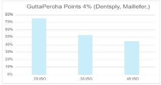
Results:
Overall, 58.9% of the GP points assessed were in accordance with the ISO size. Depending on the size (20, 30, 40 according to ISO), compliance was 75%, 52.5% and 44.4% in the group GP 4%, and 87.5%, 32.5% and 60% in the group GP 6%, respectively. The results were statistically significant in the GP 4% (p<0.0001) and GP 6% (p<0.05) groups. In both groups, standardisation of greater taper GP was most accurate at size 20. There were no statistically significant differences between the number of GP points with correct diameter between groups GP 4% and GP 6%.
Conclusions:
The study demonstrated the presence of differences between the ISO size and the actual size of the greater taper GP points. Therefore, the calibration of greater taper GP points before root canal filling should be a routine procedure.
When filling root canals with gutta-percha points (GP), good adaptation of the point to the apical diameter must be achieved through the use of a point corresponding to the size of the master apical file (MAF). GP points with a greater taper, designed to fit into root canals shaped with files with a greater taper, are currently in common use. The study was performed to evaluate the ISO compliance of GP greater taper points (4% and 6%).
Material and methods:
Two boxes of GP in sizes 20, 30 and 40 (Dentsply Maillefer, Switzerland) with a taper of 4% (GP 4% group) and 6% (GP 6% group) were used. The percentage of GPs conforming to the ISO size and those not conforming to the ISO size was determined by assessing the fit of the GP apex diameter (D0) with an endodontic gauge (Dentsply Maillefer, Switzerland). Data were analysed using the chi2 test with a significance level of 0.05.
Results:
Overall, 58.9% of the GP points assessed were in accordance with the ISO size. Depending on the size (20, 30, 40 according to ISO), compliance was 75%, 52.5% and 44.4% in the group GP 4%, and 87.5%, 32.5% and 60% in the group GP 6%, respectively. The results were statistically significant in the GP 4% (p<0.0001) and GP 6% (p<0.05) groups. In both groups, standardisation of greater taper GP was most accurate at size 20. There were no statistically significant differences between the number of GP points with correct diameter between groups GP 4% and GP 6%.
Conclusions:
The study demonstrated the presence of differences between the ISO size and the actual size of the greater taper GP points. Therefore, the calibration of greater taper GP points before root canal filling should be a routine procedure.
Gałan K, Bobryk S, Bagińska J. Size accuracy of the greater taper gutta-percha points – preliminary report. J Pre-Clin Clin Res. 2024; 18(2):
95–97. doi: 10.26444/jpccr/188142
REFERENCES (28)
1.
Alrahabi M, Zafar MS, Adanir N. Aspects of clinical malpractice in endodontics. Eur J Dent. 2019;13:450–8. https://doi. org/10.1055/s-0039-1700767.
2.
Khanna R, Handa A, Virk RK, Ghai D, et al. Clinical and Radiographic Evaluation of Procedural Errors during Preparation of Curved Root Canals with Hand and Rotary Instruments: A Randomized Clinical Study. Contemp Clin Dent. 2017;8(3):411–415. doi:10.4103/ccd.ccd_495_17.
3.
Bhandi S, Mashyakhy M, Abumelha AS, et al. Complete Obturation- Cold Lateral Condensation vs. Thermoplastic Techniques: A Systematic Review of Micro-CT Studies. Materials (Basel). 2021;14(14):4013. doi:10.3390/ma14144013.
4.
Salles AA, Cord AB, Sonnenman TS, et al. Comparative analysis of the diameter of MTwo® system gutta-percha points in relation to their corresponding instruments. RSBO Revista Sul-Brasileira de Odontologia [en linea]. 2013;10(1):49–55.
5.
Kalantar Motamedi MR, Mortaheb A, Zare Jahromi M, et al. Micro- CT evaluation of four root canal obturation techniques. Scanning. 2021;25:6632822. doi:10.1155/2021/6632822.
6.
De-Deusa G, Santos G, Monteiro IZ. Micro-CT assessment of gapcontaining areas along the gutta-percha-sealer interface in oval-shaped canals. Int Endod J. 2022;55(7):795–807. doi:10.1111/iej.13744.
7.
Alim BA, Garip Berker Y. Evaluation of different root canal filling techniques in severely curved canals by micro-computed tomography. Saudi Dent J. 2020;32(4):200–205. doi:10.1016/j.sdentj.2019.08.009.
8.
Kaul S, Kumar A, Badiyani BK. Comparison of Sealing Ability of Bioceramic Sealer, AH Plus, and GuttaFlow in Conservatively Prepared Curved Root Canals Obturated with Single-Cone Technique: An In vitro Study. J Pharm Bioallied Sci. 2021;3(1):857–S860. doi:10.4103/ jpbs.jpbs_52_21.
9.
Vishwanath V, Rao HM. Gutta-percha in endodontics – A comprehensive review of material science. J Conserv Dent. 2019;22(3):216–222. doi:10.4103/JCD.JCD_420_18.
10.
Pawińska M, Szczurko G, Łuczaj-Cepowicz E, et al. Cytotoxicity and oxidative stress induction by root canal sealers in human periodontal ligament fibroblasts: an in vitro study. Iran Endod J. 2021;16(3):164–175. https://doi.org/10.22037/iej.v....
11.
Hirai VHG, Machado R, Budziak MCL, et al. Percentage of gutta-percha-, sealer-, and void-filled areas in oval-shaped root canals obturated with different filling techniques: a confocal laser scanning microscopy study. Eur J Dent. 2020;14(1):8–12. https://doi.org/10.1055/s-0040....
12.
Manchanda S, Sardana D, Yiu CKY. A systematic review and metaanalysis of randomized clinical trials comparing rotary canal instrumentation techniques with manual instrumentation techniques in primary teeth. Int Endod J. 2020;53(3):333–353. doi:10.1111/iej.13359.
13.
Faghihian R, Amini K, Tahririan D. Rotary versus Manual Instrumentation for Root Canal Preparation in Primary Teeth: A Systematic Review and Meta-Analysis of Clinical Trials. Contemp Clin Dent. 2022;13(3):197–204. doi:10.4103/ccd.ccd_77_20.
14.
Ho ES, Chang JW, Cheung GS. Quality of root canal fillings using three gutta-percha obturation techniques. Restor Dent Endod. 2016;41(1):22–28. doi:10.5395/rde.2016.41.1.22.
15.
Bielawiec A, Bobryk S, Gałan K, et al. Evaluation of the standardisation of gutta-percha points. J Pre-Clin Clin Res. 2023;17(2):70–72. https:// doi.org/10.26444/jpccr/165877.
16.
Silva AVT, Freitas BE, Faclao CAM, et al. Evaluation of gutta-percha points standardization. IOSR Journal of Dental and Medical Sciences (IOSR-JDMS). 2019;18(9):45–47.
17.
Iványi I, Gyurkovics M, Várnagy E, et al. Comparison of guttapercha points of different brands. Fogorv. Sz. 2008;101(2):65–69.
18.
Bajaj N, Monga P, Mahajan P. Assessment of consistency in the dimension of gutta-percha cones of ProTaper Next and WaveOne with their corresponding number files. Eur J Dent. 2017;11(2):201–205. https://doi.org/10.4103/ejd.ej....
19.
Cunha RS, Fontana CE, Bueno CES. et al. Avaliação do diâmetro d0 de cones estandardizados de diferentes marcas comerciais através de régua calibradora. Revista Gaúcha de Odontologia. 2003;51(4):215–218.
20.
Cunningham KP, Walker MP, Kulild JC, et al. Variability of the diameter and taper of size #30, 0.04 gutta-percha cones. J Endod. 2006;32(11):1081–1084. https://doi.org/10.1016/j.joen....
21.
Castilho EH, Brito MLB, Borges ML, Machado ME, et al. Accuracy of the tip diameter on gutta-percha cones of different tapers Arq. Odontol. 2014;50(3):138–141. https://doi.org/10.7308/aodont....
22.
Bueno JPT, Melo TAF, Kunert GG. Evaluation of the tip of standardized D0 gutta percha cones of four Rotary systems, by means of an endodontic ruler. Rev Gaúch Odontol. 2017;65(4):299–302 doi:10.1590/1981-863720170002000023168.
23.
Moule AJ, Kellaway R, Clarkson R, et al. Variability of master gutta-percha cones. Aust Endod J. 2002;28(1):38– 43. https://doi. org/10.1111/j.1747-4477.2002.tb00365.x.
24.
American Dental Association. Council of Scientific Research. Revised American National Standard/American Dental Association Standard no. 78 For Dental Obturating Cones. 2013. https://webstore.ansi.org/ standards/ada/ansiada782013 accessed 15.02.2024.
25.
Polski Komitet Normalizacyjny. PN-ENG ISO 6877:2021 (ang.). Dentistry. Endodontic Obturating materials. (access 24.01.2023).
26.
Martínez I, Lozano A, Sanz JL, et al. Diameter and taper variability of gutta-percha cones adapted to TruNatomyTM and RotateTM rotary file systems. J Clin Exp Dent. 2023 Jan 1;15(1):e17–e22. https://doi. org/10.4317/jced.59992.
27.
Haupt F, Seidel M, Rizk M, et al. Diameter and Taper Variability of Single-file Instrumentation Systems and Their Corresponding Guttapercha Cones. J Endod. 2018;44(9):1436–1441 https://doi.org/10.1016/j. joen.2018.06.005.
28.
Dadalti MT, Ormiga F, Araújo MCP, et al. Accuracy of the initial diameter of finishing files and gutta-percha cones of the ProTaper Universal® system. Revista Científica do CRO-RJ / Rio de Janeiro Dental Journal. 2018; 3(2): 32–36. https://doi.org/10.29327/24816....
Share
RELATED ARTICLE
| eISSN: | 1898-7516 |
| ISSN: | 1898-2395 |
We process personal data collected when visiting the website. The function of obtaining information about users and their behavior is carried out by voluntarily entered information in forms and saving cookies in end devices. Data, including cookies, are used to provide services, improve the user experience and to analyze the traffic in accordance with the Privacy policy. Data are also collected and processed by Google Analytics tool (more).
You can change cookies settings in your browser. Restricted use of cookies in the browser configuration may affect some functionalities of the website.
You can change cookies settings in your browser. Restricted use of cookies in the browser configuration may affect some functionalities of the website.