Online first
About the Journal
Current issue
Archive
Publication Ethics
Anti-Plagiarism system
Instructions for Authors
Instructions for Reviewers
Editorial Office
Editorial Board
Contact
Reviewers
All Reviewers
2025
2024
2023
2022
2021
2020
2019
2018
2017
2016
General Data Protection Regulation (RODO)
RESEARCH PAPER
Evaluation of the standardisation of gutta-percha points
1
Student Research Group ‘StuDentio’, Department of Dentistry Propaedeutics, Medical University, Białystok, Poland
2
Department of Dentistry Propaedeutic, Medical University, Białystok, Poland
Corresponding author
J Pre Clin Clin Res. 2023;17(2):70-72
KEYWORDS
TOPICS
ABSTRACT
Introduction and objective:
In order to archieve good adaptation to the apical diameter, the master gutta-percha (GP) point should match the last instrument used at the working length. The aim of this study was to determine whether the diameters of the standard gutta-percha points comply with the ISO standard.
Material and methods:
The diameter at the tip (D0) of GP points (2% taper) sizes 30, 35 and 40 manufactured by Meta Biomed, South Korea (group A) and DiaDent, South Korea (group B) was assessed using an endodontic gauge (Dentsply Maillefer, Switzerland). The percentage of points larger than ISO size, compliant and smaller than ISO size, was calculated. Data were analysed using the chi2 test, with a significance level of 0.05.
Results:
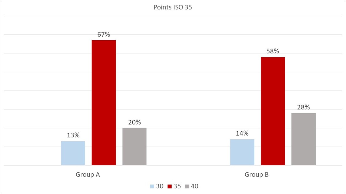
71.2% of assessed points met the ISO size requirements, 16.2% were smaller and 12.6% were larger than expected. These percentages were 76%, 12.6% and 11.4% in group A, and 66.4%, 19.6% and 14% in group B, respectively. There were no statistically significant differences between the percentage of proper and incorrect sizes in groups A and B overall, or for sizes 35 and 40. Only for group A 30 the standardisation of the points was more accurate than for group B 30 (p<0.001).
Conclusions:
This study shows that the dimensions of gutta-percha points may differ from the ISO standard. The use of an endodontic gauge can help select points with a good adaptation to the apical diameter and should be introduced as a standard procedure when obturating root canals.
In order to archieve good adaptation to the apical diameter, the master gutta-percha (GP) point should match the last instrument used at the working length. The aim of this study was to determine whether the diameters of the standard gutta-percha points comply with the ISO standard.
Material and methods:
The diameter at the tip (D0) of GP points (2% taper) sizes 30, 35 and 40 manufactured by Meta Biomed, South Korea (group A) and DiaDent, South Korea (group B) was assessed using an endodontic gauge (Dentsply Maillefer, Switzerland). The percentage of points larger than ISO size, compliant and smaller than ISO size, was calculated. Data were analysed using the chi2 test, with a significance level of 0.05.
Results:
71.2% of assessed points met the ISO size requirements, 16.2% were smaller and 12.6% were larger than expected. These percentages were 76%, 12.6% and 11.4% in group A, and 66.4%, 19.6% and 14% in group B, respectively. There were no statistically significant differences between the percentage of proper and incorrect sizes in groups A and B overall, or for sizes 35 and 40. Only for group A 30 the standardisation of the points was more accurate than for group B 30 (p<0.001).
Conclusions:
This study shows that the dimensions of gutta-percha points may differ from the ISO standard. The use of an endodontic gauge can help select points with a good adaptation to the apical diameter and should be introduced as a standard procedure when obturating root canals.
Bielawiec A, Bobryk S, Gałan K, Bagińska J, Kobus A. Evaluation of the standardisation of gutta-percha points. J Pre-Clin Clin Res. 2023; 17(2): 70–72. doi: 10.26444/jpccr/165877
REFERENCES (14)
1.
Salles AA, Cord CB, Sonnemann TS, at al. Comparative analysis of the diameter of MTwo® system gutta-percha points in relation to their corresponding instruments. RSBO (Online). 2013;10(1):49–55.
2.
Kalantar Motamedi MR, Mortaheb A, Zare Jahromi M, Gilbert BE. Micro-CT evaluation of four root canal obturation techniques. Scanning. 2021;25;2021:6632822. doi:10.1155/2021/6632822.
3.
Hirai VHG, Machado R, Budziak MCL, et al. Percentage of gutta-percha-, sealer-, and void-filled areas in oval-shaped root canals obturated with different filling techniques: a confocal laser scanning microscopy study. Eur J Dent. 2020;14(1):8–12. doi:10.1055/s-0040-1701543.067832.
4.
Pawińska M, Szczurko G, Łuczaj-Cepowicz E, et al. Cytotoxicity and oxidative stress induction by root canal sealers in human periodontal ligament fibroblasts: an in vitro study. Iran Endod J. 2021;16(3):164–175.
5.
Collado-González M, Tomás-Catalá CJ, Oñate-Sánchez RE, et al. Cytotoxicity of GuttaFlow Bioseal, GuttaFlow2, MTA Fillapex, and AH Plus on human periodontal ligament stem cells. J Endod. 2017;43(5):816–822. doi:10.1016/j.joen.2017.01.001.
6.
Kozuń A, Banaszek K, Sawicki J. Evaluation of endodontic gauge used for measurement of gutta-percha cones – preliminary report. Dent Med Probl. 2009;46(4):450–453.
7.
Bajaj N, Monga P, Mahajan P. Assessment of consistency in the dimension of gutta-percha cones of ProTaper Next and WaveOne with their corresponding number files. Eur J Dent. 2017;11(2):201–205. doi:10.4103/ejd.ejd_167_16.
8.
Moule AJ, Kellaway R, Clarkson R, et al. Variability of master gutta-percha cones. Aust Endod J. 2002;28(1):38– 43. doi:10.1111/j.1747-4477.2002.tb00365.x.
9.
Polski Komitet Normalizacyjny. PN-ENG ISO 6877:2021 (ang.). Dentistry. Endodontic Obturating materials. (access 2013.01.24).
10.
Mirmohammadi H, Sitarz M, Shemesh H. Intramanufacture diameter variability of rotary files and their corresponding gutta-percha cones using laser scan micrometre. Iran Endod J. 2018;13(2):159–162. doi:10.22037/iej.v13i2.14710. PMID:29707008; PMCID: PMC5911287.
11.
Martínez I, Lozano A, Sanz JL, et al. Diameter and taper variability of gutta-percha cones adapted to TruNatomyTM and RotateTM rotary file systems. J Clin Exp Dent. 2023;15(1):e17-e22. doi:10.4317/jced.59992.
12.
Haupt F, Seidel M, Rizk M, et al. Diameter and taper variability of single-file instrumentation systems and their corresponding gutta-percha cones. J Endod. 2018;44(9):1436–1441. doi:10.1016/j.joen.2018.06.005.
13.
Silva AVT, Freitas BE, Faclao CAM, et al. Evaluation of gutta-percha points standardization. IOSR Journal of Dental and Medical Sciences (IOSR-JDMS). 2019;18(9):45–47.
14.
Goldberg F, Massone EJ, Pruskin E, et al. SEM study of surface architecture of gutta−percha cones. Endod Dent Traumatol. 1991;7(1):15–18.
| eISSN: | 1898-7516 |
| ISSN: | 1898-2395 |
We process personal data collected when visiting the website. The function of obtaining information about users and their behavior is carried out by voluntarily entered information in forms and saving cookies in end devices. Data, including cookies, are used to provide services, improve the user experience and to analyze the traffic in accordance with the Privacy policy. Data are also collected and processed by Google Analytics tool (more).
You can change cookies settings in your browser. Restricted use of cookies in the browser configuration may affect some functionalities of the website.
You can change cookies settings in your browser. Restricted use of cookies in the browser configuration may affect some functionalities of the website.