Online first
About the Journal
Current issue
Archive
Publication Ethics
Anti-Plagiarism system
Instructions for Authors
Instructions for Reviewers
Editorial Office
Editorial Board
Contact
Reviewers
All Reviewers
2025
2024
2023
2022
2021
2020
2019
2018
2017
2016
General Data Protection Regulation (RODO)
REVIEW PAPER
Pictorial review of COVID-19 in the Lublin Region – Imaging disease progression with CXR and CT
1
2nd Department of Radiology, Medical University, Lublin, Poland
2
Department of Internal Medicine, Medical University, Lublin, Poland
Corresponding author
Ewa Kurys-Denis
2nd Department of Radiology, ul. St. Staszica 16, Medical University, 20-081 Lublin, Poland
2nd Department of Radiology, ul. St. Staszica 16, Medical University, 20-081 Lublin, Poland
J Pre Clin Clin Res. 2021;15(1):40-45
KEYWORDS
TOPICS
ABSTRACT
Introduction:
COVID-19 is a disease caused by SARS-Cov-2 that has reached the pandemic status and has infected in one year more than 62 million people. Clinical symptoms range from barely noticeable to very severe. It is crucial to recognize imaging patterns of COVID-19, allowing for better diagnosis and treatment. Diagnostic imaging is also essential in monitoring patients in the course of the disease.
Objective:
In our pictorial review we describe the most common pulmonary manifestations of COVID-19, and show the typical and non-typical features of COVID-19 encountered in our hospital in Lublin, Poland. Imaging the disease progression is also visualized to help realize how pulmonary changes occur over the time.
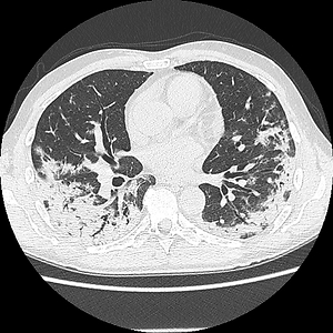
State of knowledge and Conclusions:
COVID-19 involves both lung parenchyma and interstitium and has multiple imaging features, varying form ground glass opacities (GGO), consolidations, reticular interstitial pattern, honeycombing or crazy-paving. Mediastinal and hilar lymph node enlargement or pleural effusion may appear, but are rare and atypical. GGO are located peripherally, bilaterally and predominantly in the lower lobes, and in the early stage are better seen on CT imaging. Progression of imaging findings take different times, with the peak of of imaging features appearing around 10–14 days after initial symptoms. While it is harder to discern subtle changes on CXR, progression can be very well monitored by his method. Final pulmonary consequences of the disease should be assessed with the use of CT.
COVID-19 is a disease caused by SARS-Cov-2 that has reached the pandemic status and has infected in one year more than 62 million people. Clinical symptoms range from barely noticeable to very severe. It is crucial to recognize imaging patterns of COVID-19, allowing for better diagnosis and treatment. Diagnostic imaging is also essential in monitoring patients in the course of the disease.
Objective:
In our pictorial review we describe the most common pulmonary manifestations of COVID-19, and show the typical and non-typical features of COVID-19 encountered in our hospital in Lublin, Poland. Imaging the disease progression is also visualized to help realize how pulmonary changes occur over the time.
State of knowledge and Conclusions:
COVID-19 involves both lung parenchyma and interstitium and has multiple imaging features, varying form ground glass opacities (GGO), consolidations, reticular interstitial pattern, honeycombing or crazy-paving. Mediastinal and hilar lymph node enlargement or pleural effusion may appear, but are rare and atypical. GGO are located peripherally, bilaterally and predominantly in the lower lobes, and in the early stage are better seen on CT imaging. Progression of imaging findings take different times, with the peak of of imaging features appearing around 10–14 days after initial symptoms. While it is harder to discern subtle changes on CXR, progression can be very well monitored by his method. Final pulmonary consequences of the disease should be assessed with the use of CT.
Kurys-Denis E, Podgórska K, Prystupa A. Pictorial review of COVID-19 from the Lublin region. Imaging disease progression with CXR and CT.
J Pre-Clin Clin Res. 2021; 15(1): 40–45. doi: 10.26444/aaem/132452
REFERENCES (31)
2.
Lotfi M, Hamblin MR, Rezaei N. COVID-19: Transmission, prevention, and potential therapeutic opportunities. Clin Chim Acta. 2020 Sep; 508: 254–266. https://doi.org/10.1016/j.cca.....
3.
Zhang G, Hu C, Luo L, et al. Clinical features and short-term outcomes of 221 patients with COVID-19 in Wuhan, China. J Clin Virol. 2020 Jun; 127: 104364. https://doi.org/10.1016/j.jcv.....
4.
Zhou S, Wang Y, Zhu T, et al. CT Features of Coronavirus Disease 2019 (COVID-19) Pneumonia in 62 Patients in Wuhan, China. AJR Am J Roentgenol. 2020 Jun; 214(6): 1287–1294. https://doi.org/10.2214/AJR.20....
5.
Zhou S, Wang Y, Zhu T, et al. CT Features of Coronavirus Disease 2019 (COVID-19) Pneumonia in 62 Patients in Wuhan, China. AJR Am J Roentgenol. 2020 Jun; 214(6): 1287–1295. https://doi.org/10.2214/AJR.20....
6.
Gao JW, Rizzo S, Ma LH, et al. Written on behalf of the AME Lung Cancer Collaborative Group. Pulmonary ground-glass opacity: computed tomography features, histopathology and molecular pathology. Transl Lung Cancer Res. 2017 Feb; 6(1): 68–75. https://doi.org/10.21037/tlcr.....
7.
Collins J, Stern EJ. Ground-glass opacity at CT: the ABCs. AJR Am J Roentgenol. 1997 Aug; 169(2): 355–67. https://doi.org/10.2214/ajr.16....
8.
Ding X, Xu J, Zhou J, et al. Chest CT findings of COVID-19 pneumonia by duration of symptoms. Eur J Radiol. 2020 Jun; 127: 10900.
9.
https://doi.org/10.1016/j.ejra.... Abbasi-Oshaghi E, Mirzaei F, Farahani F, et al. Diagnosis and treatment of coronavirus disease 2019 (COVID-19): Laboratory, PCR, and chest CT imaging findings. Int J Surg. 2020 Jul; 79: 143–153. https://doi.org/10.1016/j.ijsu....
10.
Rodriguez-Morales AJ, Cardona-Ospina JA, Gutiérrez-Ocampo E, et al. Latin American Network of Coronavirus Disease 2019-COVID-19 Research (LANCOVID-19). Electronic address: https://www.lancovid.org. Clinical, laboratory and imaging features of COVID-19: A systematic review and meta-analysis. Travel Med Infect Dis. 2020 Mar-Apr; 34: 101623. https://doi.org/10.1016/j.tmai....
11.
Hu Q, Guan H, Sun Z, et al. Early CT features and temporal lung changes in COVID-19 pneumonia in Wuhan, China. Eur J Radiol. 2020 Jul; 128: 109017. https://doi.org/10.1016/j.ejra....
12.
Wang D, Hu B, Hu C, et al. Clinical Characteristics of 138 Hospitalized Patients With 2019 Novel Coronavirus-Infected Pneumonia in Wuhan, China. JAMA. 2020 Mar 17; 323(11): 1061–1069. http://doi.org/10.1001/jama.20....
13.
Siordia JA Jr. Epidemiology and clinical features of COVID-19: A review of current literature. J Clin Virol. 2020 Jun; 127: 104357. https://doi.org/10.1016/j.jcv.....
14.
Wan S, Li M, Ye Z, et al. CT Manifestations and Clinical Characteristics of 1115 Patients with Coronavirus Disease 2019 (COVID-19): A Systematic Review and Meta-analysis. Acad Radiol. 2020 Jul; 27(7): 910–921. https://doi.org/10.1016/j.acra....
15.
Neji H, Attia M, Affes M, et al. Interstitial lung diseases: Imaging contribution to diagnosis and elementary radiological lesions. Semin Diagn Pathol. 2018 Sep; 35(5): 297–303. https://doi.org/10.1053/j.semd....
16.
Fu F, Lou J, Xi D, et al. Chest computed tomography findings of coronavirus disease 2019 (COVID-19) pneumonia. Eur Radiol. 2020 Oct; 30(10): 5489–5498. htpps://doi.org/10.1007/s00330-020-06920-8.
17.
Nicola M, O’Neill N, Sohrabi C, et al. Evidence based management guideline for the COVID-19 pandemic – Review article. Int J Surg. 2020 May; 77: 206–216. http://doi.org/10.1016/j.ijsu.....
18.
Liu J, Chen T, Yang H, et al. Clinical and radiological changes of hospitalised patients with COVID-19 pneumonia from disease onset to acute exacerbation: a multicentre paired cohort study. Eur Radiol. 2020 Oct; 30(10): 5702–5708. https://doi.org/10.1007/s00330....
19.
Li X, Fang X, Bian Y, et al. Comparison of chest CT findings between COVID-19 pneumonia and other types of viral pneumonia: a two-center retrospective study. Eur Radiol. 2020 Oct; 30(10): 5470–5478. https://doi.org/10.1007/s00330....
20.
Fang Y, Zhang H, Xie J, et al. Sensitivity of Chest CT for COVID-19: Comparison to RT-PCR. Radiology. 2020 Aug; 296(2): E115-E117. https://doi.org/10.1148/radiol....
21.
Kanne JP, Little BP, Chung JH, et al. Essentials for Radiologists on COVID-19: An Update-Radiology Scientific Expert Panel. Radiology. 2020 Aug; 296(2): E113-E114. https://doi.org/10.1148/radiol....
22.
Kooraki S, Hosseiny M, Myers L, et al. Coronavirus (COVID-19) Outbreak: What the Department of Radiology Should Know. J Am Coll Radiol. 2020 Apr; 17(4): 447–451. https://doi.org/10.1016/j.jacr....
23.
Jacobi A, Chung M, Bernheim A, et al. Portable chest X-ray in coronavirus disease-19 (COVID-19): A pictorial review. Clin Imaging. 2020 Aug; 64: 35–42. https://doi.org/10.1016/j.clin....
24.
Salehi S, Abedi A, Balakrishnan S, et al. Coronavirus Disease 2019 (COVID-19): A Systematic Review of Imaging Findings in 919 Patients. AJR Am J Roentgenol. 2020 Jul; 215(1): 87–93. https://doi.org/10.2214/AJR.20....
25.
Jajodia A, Ebner L, Heidinger B, Chaturvedi A, Prosch H. Imaging in corona virus disease 2019 (COVID-19)-A Scoping review. Eur J Radiol Open. 2020 May 11; 7: 100237. https://doi.org/10.1016/j.ejro....
26.
Pan F, Ye T, Sun P, et al. Time Course of Lung Changes at Chest CT during Recovery from Coronavirus Disease 2019 (COVID-19). Radiology. 2020 Jun; 295(3): 715–721. https://doi.org/10.1148/radiol....
27.
Wong HYF, Lam HYS, Fong AH, et al. Frequency and Distribution of Chest Radiographic Findings in Patients Positive for COVID-19. Radiology. 2020 Aug; 296(2): E72-E78. https://doi.org/10.1148/radiol....
28.
Guan CS, Lv ZB, Yan S, et al. Imaging Features of Coronavirus disease 2019 (COVID-19): Evaluation on Thin-Section CT. Acad Radiol. 2020 May; 27(5): 609–613. https://doi.org/10.1016/j.acra....
29.
Wu G, Li X. Mobile X-rays are highly valuable for critically ill COVID patients. Eur Radiol. 2020 Sep; 30(9): 5217–5219. https://doi.org/10.1007/s00330....
30.
Simpson S, Kay FU, Abbara S, et al. Radiological Society of North America Expert Consensus Statement on Reporting Chest CT Findings Related to COVID-19. Endorsed by the Society of Thoracic Radiology, the American College of Radiology, and RSNA – Secondary Publication. J Thorac Imaging. 2020 Jul; 35(4): 219–227. https://doi.org/10.1097/RTI.00....
Share
RELATED ARTICLE
| eISSN: | 1898-7516 |
| ISSN: | 1898-2395 |
We process personal data collected when visiting the website. The function of obtaining information about users and their behavior is carried out by voluntarily entered information in forms and saving cookies in end devices. Data, including cookies, are used to provide services, improve the user experience and to analyze the traffic in accordance with the Privacy policy. Data are also collected and processed by Google Analytics tool (more).
You can change cookies settings in your browser. Restricted use of cookies in the browser configuration may affect some functionalities of the website.
You can change cookies settings in your browser. Restricted use of cookies in the browser configuration may affect some functionalities of the website.