Online first
About the Journal
Current issue
Archive
Publication Ethics
Anti-Plagiarism system
Instructions for Authors
Instructions for Reviewers
Editorial Office
Editorial Board
Contact
Reviewers
All Reviewers
2025
2024
2023
2022
2021
2020
2019
2018
2017
2016
General Data Protection Regulation (RODO)
CASE REPORT
Radiological features of perivascular epithelioid cell tumours (PEComas) in a paediatric patient – Case report
1
Medical University, Lublin, Poland
2
Medical University of Lublin, Poland
J Pre Clin Clin Res. 2022;16(1):9-12
KEYWORDS
TOPICS
ABSTRACT
Introduction:
Perivascular epithelioid cell tumour (PEComa) is a rare family of mesenchymal tumours composed of epithelioid cells. Due to its very rare occurrence, little information is available on the imaging characteristics of this type of lesion.
Case Report:
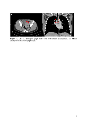
A 16-year-old patient was admitted for diagnosis of an incidentally found lesion of unclear origin on MRI of the lumbar spine. Targeted MRI revealed a pathological solid-cystic mass within the pelis, in the left rectovesical pouch, infiltrating the bladder wall. A biopsy of the tumour was performed. On histopathological examination, a PEComa tumour was diagnosed. Control examinations were performed. The tumour was treated with embolization, surgical resection and Sirolimus therapy.
Conclusions:
The tumour showed the characteristic features of PEC-oma on imaging studies reported in the literature. Radiological diagnosis is not fully specific, therefore histopathological examination is necessary for a definitive diagnosis
Perivascular epithelioid cell tumour (PEComa) is a rare family of mesenchymal tumours composed of epithelioid cells. Due to its very rare occurrence, little information is available on the imaging characteristics of this type of lesion.
Case Report:
A 16-year-old patient was admitted for diagnosis of an incidentally found lesion of unclear origin on MRI of the lumbar spine. Targeted MRI revealed a pathological solid-cystic mass within the pelis, in the left rectovesical pouch, infiltrating the bladder wall. A biopsy of the tumour was performed. On histopathological examination, a PEComa tumour was diagnosed. Control examinations were performed. The tumour was treated with embolization, surgical resection and Sirolimus therapy.
Conclusions:
The tumour showed the characteristic features of PEC-oma on imaging studies reported in the literature. Radiological diagnosis is not fully specific, therefore histopathological examination is necessary for a definitive diagnosis
Karska K, Kozioł I, Leśniewska M, Budzyńska J, Woźniak M. Radiological features of perivascular epithelioid cell tumours (PEComas) in
a paediatric patient – Case report. J Pre-Clin Clin Res. 2022; 16(1): 9–12. doi: 10.26444/jpccr/146536
REFERENCES (10)
1.
Tan Y, Zhang H, Xiao EH. Perivascular epithelioid cell tumour: dynamic CT, MRI and clinicopathological characteristics–analysis of 32 cases and review of the literature. Clin Radiol. 2013 Jun; 68(6): 555–61. doi: 10.1016/j.crad.2012.10.021. Epub 2012 Dec 11. PMID: 23245276.
2.
Diestelkamp T, Mikes Z, Wilson-Smith R, Germaine P. Radiological findings of two neoplasms with perivascular epithelioid cell differentiation. Radiol Case Rep. 2017 Aug 19; 12(4): 845–849. doi: 10.1016/j.radcr.2017.06.001 PMID: 29484084; PMCID: PMC5823291.
3.
Handa A, Fujita K, Kono T, Komori K, Hirobe S, Fukuzawa R. Radiological findings of perivascular epithelioid cell tumour (PEComa) of the falciform ligament. J Med Imaging Radiat Oncol. 2016 Dec; 60(6): 741–743. doi: 10.1111/1754-9485.12460 Epub 2016 Apr 20. PMID: 27094767.
4.
Gkizas CV, Tsili AC, Katsios C, Argyropoulou MI. Perirenal PEComa: Computed Tomography Findings and Differential Diagnosis. J Clin Imaging Sci. 2015 Dec 31; 5: 69. doi: 10.4103/2156-7514.172977. PMID: 26900493; PMCID: PMC4736062.
5.
Zhao J, Teng H, Zhao R, Ding W, Yu K, Zhu L, Zhang J, Han Y. Malignant perivascular epithelioid cell tumour of the lung synchronous with a primary adenocarcinoma: one case report and review of the literature. BMC Cancer. 2019 Mar 15; 19(1): 235. doi:10.1186/s12885-019-5383-0 PMID: 30876389; PMCID: PMC6419825.
6.
Tirumani SH, Shinagare AB, Hargreaves J, Jagannathan JP, Hornick JL, Wagner AJ, Ramaiya NH. Imaging features of primary and metastatic malignant perivascular epithelioid cell tumours. AJR Am J Roentgenol. 2014 Feb; 202(2): 252–8. doi: 10.2214/AJR.13.10909 PMID: 24450662.
7.
Xuesong D, Hong G, Weiguo Z. Bladder Perivascular Epithelioid Cell Tumour: Dynamic CT and MRI Presentation of 2 Cases With 2-year Follow-up and Review of the Literature. Clin Genitourin Cancer. 2019 Oct; 17(5): e916-e922. doi: 10.1016/j.clgc.2019.06.016 Epub 2019 Jul 2. PMID: 31327725.
8.
Faria SC, Elsherif SB, Sagebiel T, Cox V, Rao B, Lall C, Bhosale PR. Ischiorectal fossa: benign and malignant neoplasms of this “ignored” radiological anatomical space. Abdom Radiol (NY). 2019 May; 44(5): 1644–1674. doi: 10.1007/s00261-019-01930-7 PMID: 30955068.
9.
Musella A, De Felice F, Kyriacou AK, Barletta F, Di Matteo FM, Marchetti C, Izzo L, Monti M, Benedetti Panici P, Redler A, D’Andrea V. Perivascular epithelioid cell neoplasm (PEComa) of the uterus: A systematic review. Int J Surg. 2015 Jul; 19: 1–5. doi: 10.1016/j. ijsu.2015.05.002 Epub 2015 May 14. PMID: 25981307.
10.
Nishio N, Kido A, Minamiguchi S, Kiguchi K, Kurata Y, Nakao KK, Kuwahara R, Yajima R, Otani S, Mandai M, Togashi K, Minami M.MR findings of uterine PEComa in patients with tuberous sclerosis: report of two cases. Abdom Radiol (NY). 2019 Apr; 44(4): 1256–1260. doi: 10.1007/s00261-019-01918-3 PMID: 30778737.
Share
RELATED ARTICLE
| eISSN: | 1898-7516 |
| ISSN: | 1898-2395 |
We process personal data collected when visiting the website. The function of obtaining information about users and their behavior is carried out by voluntarily entered information in forms and saving cookies in end devices. Data, including cookies, are used to provide services, improve the user experience and to analyze the traffic in accordance with the Privacy policy. Data are also collected and processed by Google Analytics tool (more).
You can change cookies settings in your browser. Restricted use of cookies in the browser configuration may affect some functionalities of the website.
You can change cookies settings in your browser. Restricted use of cookies in the browser configuration may affect some functionalities of the website.