Online first
About the Journal
Current issue
Archive
Publication Ethics
Anti-Plagiarism system
Instructions for Authors
Instructions for Reviewers
Editorial Office
Editorial Board
Contact
Reviewers
All Reviewers
2025
2024
2023
2022
2021
2020
2019
2018
2017
2016
General Data Protection Regulation (RODO)
RESEARCH PAPER
Morphological assessment of the incisive canal using cone beam computed tomography in a Polish population sample
1
Medical University, Lublin, Poland
J Pre Clin Clin Res. 2024;18(2):103-108
KEYWORDS
TOPICS
ABSTRACT
Introduction and objective:
The nasopalatine canal (NPC) connects the floor of the nasal cavity with the anterior part of the hard palate. The anatomical variability of the NPC dimensions and the thickness of maxillary bone anterior to the NPC is related to age, gender and ethnicity. Also the shape, position and number of NPC foramina varies in different populations. Nowadays, cone beam computed tomography (CBCT) is often used in dentistry and contributes to the depiction of anatomical landmarks. The aim of the study is analysis of the nasopalatine canal morphology in CBCT examinations in a sample of the Polish population.
Material and methods:
One hundred consecutive CBCT scans were analyzed. The studied group included 55 women and 45 men, age range: 20 – 30 years. The following criteria were recorded: shape, length and width in the narrowest part of the canal, antero-posterior measurement of nasal and palatal foramina, and medio-lateral diameter of incisive foramen, number of foramina of Stenson and division of NPC, presence of canalis sinuosus and thickness of maxillary bone anterior to the NPC.
Results:
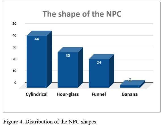
The NPC measurements were gender-related with mean values higher in males. There was no correlation according to gender in shape of the canal, presence of Stenson’s foramina and canalis sinuosus. The most common was cylindrical shape, and the rarest a banana-like shape. The frequency of single NPC canal was the highest and more often observed in females.
Conclusions:
It was found that the morphology of the NPC varied in the sample of the population, which highlights the importance of preoperative imaging diagnosis
The nasopalatine canal (NPC) connects the floor of the nasal cavity with the anterior part of the hard palate. The anatomical variability of the NPC dimensions and the thickness of maxillary bone anterior to the NPC is related to age, gender and ethnicity. Also the shape, position and number of NPC foramina varies in different populations. Nowadays, cone beam computed tomography (CBCT) is often used in dentistry and contributes to the depiction of anatomical landmarks. The aim of the study is analysis of the nasopalatine canal morphology in CBCT examinations in a sample of the Polish population.
Material and methods:
One hundred consecutive CBCT scans were analyzed. The studied group included 55 women and 45 men, age range: 20 – 30 years. The following criteria were recorded: shape, length and width in the narrowest part of the canal, antero-posterior measurement of nasal and palatal foramina, and medio-lateral diameter of incisive foramen, number of foramina of Stenson and division of NPC, presence of canalis sinuosus and thickness of maxillary bone anterior to the NPC.
Results:
The NPC measurements were gender-related with mean values higher in males. There was no correlation according to gender in shape of the canal, presence of Stenson’s foramina and canalis sinuosus. The most common was cylindrical shape, and the rarest a banana-like shape. The frequency of single NPC canal was the highest and more often observed in females.
Conclusions:
It was found that the morphology of the NPC varied in the sample of the population, which highlights the importance of preoperative imaging diagnosis
ACKNOWLEDGEMENTS
CBCT – cone beam computed tomography; IC – incisive canal; FOV – field of view; NPC – nasopalatine canal
Piskórz M, Kiełt W, Kozłowska J, Futyma-Gąbka K, Różyło-Kalinowska I. Morphological assessment of the incisive canal using cone beam
computed tomography – in Polish population sample. J Pre-Clin Clin Res. 2024; 18(2): 103–108. doi: 10.26444/jpccr/186527
REFERENCES (25)
1.
Görürgöz C, Öztaş B. Anatomic characteristics and dimensions of the nasopalatine canal: a radiographic study using cone-beam computed tomography. Folia Morphol (Warsz). 2021;80(4):923–934.
2.
Nasseh I, Aoun G, Sokhn S. Assessment of the nasopalatine canal: An anatomical study. Acta Inform Med. 2017;25(1):34–38.
3.
Friedrich RE, Laumann F, Zrnc T, Assaf AT. The Nasopalatine Canal in Adults on Cone Bean Computed Tomograms – A Clinical Study and Review of the Literature. In Vivo. 2015;29(4):467–486.
4.
Lake S, Iwanaga J, Kikuta S, Oskouian RJ, Loukas M, Tubbs RS. The Incisive Canal: A Comprehensive Review. Cureus. 2018;10(7):e3069.
5.
Bahsi I, Orhan M, Kervancioglu P, Yalçın ED, Aktan AM. Anatomical evaluation of nasopalatine canal on cone beam computed tomography images. Folia Morphol (Warsz). 2019;78(1):153–162.
6.
de Mello JS, Faot F, Correa G, Chagas Júnior OL. Success rate and complications associated with dental implants in the incisive canal region: a systematic review. Int J Oral Maxillofac Surg. 2017;46(12):1584–1591.
7.
Jain S, Choudhary K, Nagi R, Shukla S, Kaur N, Grover D. New evolution of cone-beam computed tomography in dentistry: Combining digital technologies. Imaging Sci Dent. 2019;49(3):179–190.
8.
Acar B, Kamburoğlu K. Morphological and volumetric evaluation of the nasopalatinal canal in a Turkish population using cone-beam computed tomography. Surg Radiol Anat. 2015;37(3):259–265.
9.
Kaasalainen T, Ekholm M, Siiskonen T, Kortesniemi M. Dental cone beam CT: An updated review. Phys Med. 2021;88:193–217.
10.
Tözüm TF, Güncü GN, Yıldırım YD, Yılmaz HG, Galindo-Moreno P, Velasco-Torres M, Al-Hezaimi K, Al-Sadhan R, Karabulut E, Wang H-L. Evaluation of Maxillary Incisive Canal Characteristics Related toDental Implant Treatment With Computerized Tomography:A ClinicalMulticenter Study. J Periodontol. 2012;83(3):337–343.
11.
Al-Ghurabi ZH, Al-Bahrani ZM. Radiographic Assessment of Nasopalatine Canal Using Cone Beam Computed Tomography. J Craniofac Surg. 2020;31(1):e4-e6.
12.
Gil-Marques B, Sanchis-Gimeno JA, Brizuela-Velasco A, Perez-Bermejo M, Larrazábal-Morón C. Differences in the shape and direction-course of the nasopalatine canal among dentate, partially edentulous and completely edentulous subjects. Anat Sci Int. 2020;95(1):76–84.
13.
Panda M, Shankar T, Raut A, Dev S, Kar AK, Hota S. Cone beam computerized tomography evaluation of incisive canal and anterior maxillary bone thickness for placement of immediate implants. J Indian Prosthodont Soc. 2018;18(4):356–363.
14.
Jayasinghe RM, Hettiarachchi PVKS, Fonseka MCN, Nanayakkara D, Jayasighe RD. Morphometric analysis of nasopalatine foramen in Sri Lankan population using CBCT. J Oral Biol Craniofac Res. 2020;10(2):238–240.
15.
Soumya P, Koppolu P, Pathakota KR, Chappidi V. Maxillary Incisive Canal Characteristics: A Radiographic Study Using Cone Beam Computerized Tomography. Radiol Res Pract. 2019;27:1–5.
16.
Mardinger O, Namani-Sadan N, Chaushu G, Schwartz-Arad D. Morphologic Changes of the Nasopalatine Canal Related to Dental Implanta-tion: A Radiologic Study in Different Degrees of Absorbed Maxillae. J Periodontol. 2008;79(9):1659–1662.
17.
Liang X, Jacobs R, Martens W, Hu Y, Adriaensens P, Quirynen M, Lambrichts I. Macro- and micro-anatomical, histological and computed tomography scan characterization of the nasopalatine canal. J Clin Periodontol. 2009;36(7):598–603.
18.
Etoz M, Sisman Y. Evaluation of the nasopalatine canal and variations with cone-beam computed tomography. Surg Radiol Anat. 2014;36(8):805–812.
19.
Sarna K, Estreed MA, Sonigra KJ, Amuti T, Opondo F, Kamau M, Ngeow WC. Anatomical Patterns of the Nasopalatine Canal and Incisive Foramen in an African Setting: A Cross-Sectional Study. Craniomaxillofac Trauma Reconstr. 2023;16(3):222–233.
20.
Kajan ZD, Kia J, Motevasseli S, Rezaian SR. Evaluation of the nasopalatine canal with cone-beam computed tomography in an Iranian population. Dent Res J (Isfahan). 2015;12(1):14–9.
21.
Safi Y, Moshfeghi M, Rahimian S, Kheirkhahi M, Manouchehri ME. Assessment of nasopalatine canal anatomic variations using cone beam computed tomography in a group of Iranian population. Iran J Radiol. 2017;14(1):e13480.
22.
Hakbilen S, Magat G. Evaluation of anatomical and morphological characteristics of the nasopalatine canal in a Turkish population by cone beam computed tomography. Folia Morphol (Warsz). 2018;77(3):527– 535.
23.
Rai S, Misra D, Misra A, Khatri M, Kidwai S, Bisla S, Jain P. Significance of morphometric and anatomic variations of nasopalatine canal on cone- beam computed tomography in anterior functional zone – A retrospective study. Ann Maxillofac Surg. 2021;11(1):108–114.
24.
Al-Amery SM, Nambiar P, Jamaludin M, John J, Ngeow WC. Cone beam computed tomography assessment of the maxillary incisive canal and foramen: Considerations of anatomical variations when placing immediate implants. PLoS One. 2015;10(2):e0117251.
25.
Kim YT, Lee JH, Jeong SN. Three-dimensional observations of the incisive foramen on cone-beam computed tomography image analysis. J Periodontal Implant Sci. 2020;50(1):48–55.
| eISSN: | 1898-7516 |
| ISSN: | 1898-2395 |
We process personal data collected when visiting the website. The function of obtaining information about users and their behavior is carried out by voluntarily entered information in forms and saving cookies in end devices. Data, including cookies, are used to provide services, improve the user experience and to analyze the traffic in accordance with the Privacy policy. Data are also collected and processed by Google Analytics tool (more).
You can change cookies settings in your browser. Restricted use of cookies in the browser configuration may affect some functionalities of the website.
You can change cookies settings in your browser. Restricted use of cookies in the browser configuration may affect some functionalities of the website.