Online first
About the Journal
Current issue
Archive
Publication Ethics
Anti-Plagiarism system
Instructions for Authors
Instructions for Reviewers
Editorial Office
Editorial Board
Contact
Reviewers
All Reviewers
2025
2024
2023
2022
2021
2020
2019
2018
2017
2016
General Data Protection Regulation (RODO)
RESEARCH PAPER
Analysis of upper airway measurements and cephalometric measurements using two-dimensional Cephalometry and threedimensional Computed Tomography in patients with skeletal Class III malocclusion.
1
Department and Clinic of Maxillofacial Surgery, Medical University, Lublin, Poland
2
Department of Orthodontics, Medical University, Lublin, Poland
3
Department of Medical Radiology II, Medical University, Lublin, Poland
Corresponding author
Remigiusz Czerkies
Medical University of Lublin Department and Clinic of Maxillofacial Surgery, Staszica 11, 20-081, Lublin, Poland
Medical University of Lublin Department and Clinic of Maxillofacial Surgery, Staszica 11, 20-081, Lublin, Poland
J Pre Clin Clin Res. 2023;17(4):225-230
KEYWORDS
TOPICS
ABSTRACT
Introduction and objective:
Skeletal Class III malocclusion, characterized by complex craniofacial irregularities, often necessitates orthognathic interventions for its resolution. The aims of the study were 1) to comprehensively assess the dimensions of the upper airway and cephalometric measurement, 2) to evaluate the values of upper airway dimensions in a three-dimensional context, in correlation with cephalometric measurements acquired in a two-dimensional format among patients afflicted with skeletal Class III malocclusion.
Material and methods:
Medical records were analysed of 18 patients diagnosed with skeletal Class III malocclusion undergoing combined orthodontic-surgical treatment. Cephalometric measurements were extracted from lateral cephalometric radiographs, and upper airway dimensions comprehensively evaluated using multi-slice spiral computed tomography scans and OsiriX software. Based on the results statistical analysis was performed.
Results:
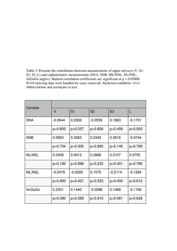
No statistically significant correlations was found between the cephalometric measurements using two-dimensional cephalometry and the upper airway dimensions using three-dimensional computed tomography in patients with skeletal Class III malocclusion.
Conclusions:
No direct influence of skeletal Class III malocclusion on upper airway dimensions was observed in study participants. Incorporating additional factors, such as soft tissue characteristics and functional aspects, may provide a more comprehensive understanding of the relationship between skeletal malocclusion and upper airway dimensions.
Skeletal Class III malocclusion, characterized by complex craniofacial irregularities, often necessitates orthognathic interventions for its resolution. The aims of the study were 1) to comprehensively assess the dimensions of the upper airway and cephalometric measurement, 2) to evaluate the values of upper airway dimensions in a three-dimensional context, in correlation with cephalometric measurements acquired in a two-dimensional format among patients afflicted with skeletal Class III malocclusion.
Material and methods:
Medical records were analysed of 18 patients diagnosed with skeletal Class III malocclusion undergoing combined orthodontic-surgical treatment. Cephalometric measurements were extracted from lateral cephalometric radiographs, and upper airway dimensions comprehensively evaluated using multi-slice spiral computed tomography scans and OsiriX software. Based on the results statistical analysis was performed.
Results:
No statistically significant correlations was found between the cephalometric measurements using two-dimensional cephalometry and the upper airway dimensions using three-dimensional computed tomography in patients with skeletal Class III malocclusion.
Conclusions:
No direct influence of skeletal Class III malocclusion on upper airway dimensions was observed in study participants. Incorporating additional factors, such as soft tissue characteristics and functional aspects, may provide a more comprehensive understanding of the relationship between skeletal malocclusion and upper airway dimensions.
Czerkies R, Kuźniarz K, Tomaszewski T, Lasota A, Krupski W. Analysis of Upper Airway Measurements and Cephalometric Measurements Using
Two-Dimensional Cephalometry and Three-Dimensional Computed Tomography in Patients with Skeletal Class III Malocclusion. J Pre-Clin
Clin Res. 2023; 17(4): 225–230. doi: 10.26444/jpccr/175314
REFERENCES (15)
1.
Martino F, Peña M, Joubert R. Surgical-orthodontic retreatment of a severe skeletal Class III malocclusion following an orthodontic camouflage. Dental Press J Orthod. 2021 Sep 10;26(4):e2119247. doi: 10.1590/2177-6709.26.4.e2119247.oar. PMID: 34524377; PMCID: PMC8439190.
2.
Eslami S, Faber J, Fateh A, et al. Treatment decision in adult patients with class III malocclusion: surgery versus orthodontics. Prog Orthod. 2018 Aug 2;19(1):28. doi:10.1186/s40510-018-0218-0. PMID: 30069814; PMCID: PMC6070451.
3.
Proffit WR, White RP, Sarver DM, Contemporary treatment of dentofacial deformity, St. Louis, Mo: Mosby; 2003.
4.
Lee CH, Park HH, Seo BM, Lee SJ. Modern trends in Class III orthognathic treatment: A time series analysis. Angle Orthod. 2017. Lee CH, Park HH, Seo BM, Lee SJ. Modern trends in Class III orthognathic treatment: A time series analysis. Angle Orthod. 2017.
5.
On SW, Kim HJ, Cho DH, et al. Silent Changes in Sleep Quality Following Mandibular Setback Surgery in Patients with Skeletal Class III Malocclusion: A Prospective Study. Sci Rep. 2019;9(1):9737. Published 2019 Jul 5, doi:10.1038/s41598-019-46166-z.
6.
Hong JS, Park YH, Kim YJ, et al. Three-dimensional changes in pharyngeal airway in skeletal class III patients undergoing orthognathic surgery, J Oral Maxillofac Surg. 2011 Nov;69(11):e401–8. doi:10.1016/j. joms.2011.02.011. Epub 2011 May 14. PubMed PMID: 21571419.
7.
Ahmed M, Shaikh A, Fida M. Diagnostic validity of different cephalometric analyses for assessment of the sagittal skeletal pattern. Dental Press J Orthod. 2018 Sep-Oct;23(5):75–81. doi:10.1590/2177- 6709.23.5.075-081.oar. PMID: 30427496; PMCID: PMC6266314.
8.
Alassiry AM. Accuracy of different cephalometric analyses in the diagnosis of class III malocclusion in Saudi and Yemeni population. J Orthod Sci. 2020 Aug 18;9:14. doi:10.4103/jos.JOS_21_20. PMID: 33354540; PMCID: PMC7749450.
9.
Bastir M, Megía I, Torres-Tamayo N, et al. Three-dimensional analysis of sexual dimorphism in the soft tissue morphology of the upper airways in a human population. Am J Phys Anthropol. 2020 Jan;171(1):65–75. doi:10.1002/ajpa.23944. PMID: 31837016.
10.
Alfawzan AA. Assessment of airway dimensions in skeletal Class I malocclusion patients with various vertical facial patterns: A cephalometric study in a sample of the Saudi population. J Orthod Sci. 2020 Aug 18;9:12. https://doi.org/10.4103/jos.JO....
11.
Paul D, Varma S, Ajith VV. Airway in Class I and Class II skeletal pattern: A computed tomography study. Contemp Clin Dent. 2015 Jul-Sep;6(3):293–8. https://doi.org/10.4103/0976-2....
12.
Aby DM, Sagarkar RM, Mathew S, et al. Comparison of Airway Morphology and Volume in Skeletal Class I and Class II Patients.Using Cone-beam Computed Tomography: A Cross-sectional Study. World J Dent. 2020;11(5):380–385. https://www.wjoud. com/abstractArticleContentBrowse/WJOUD/21902/JPJ/fullText. doi:10.5005/jp-journals-10015-1754.
13.
Emsaeili F, Sadrhaghighi A, Sadeghi-Shabestar, et al. Comparison of superior airway dimensions and cephalometric anatomic landmarks between 8–12-year-old children with obstructive sleep apnea and healthy children using CBCT images. J Dent Res Dent Clin Dent Prospects. 2022 Winter;16(1):18–23. doi:10.34172/joddd.2022.003. Epub 2022 May 29. PMID: 35936930; PMCID: PMC9339744.
14.
Neelapu BC, Kharbanda OP, Sardana HK, Balachandran R, Sardana V, Kapoor P, Gupta A, Vasamsetti S. Craniofacial and upper airway morphology in adult obstructive sleep apnea patients: A systematic review and meta-analysis of cephalometric studies. Sleep Med Rev. 2017 Feb;31:79–90. doi:10.1016/j.smrv.2016.01.007. Epub 2016 Jan 30. PMID: 27039222.
15.
Campos A, Cebola P, Simões Dias S, Pais JP, Sousa S, Cardoso S, Paço J, Caroça C. Upper airway assessment in obstructive sleep apnea patients: can computed tomography with lateral cephalometry replace druginduced sleep endoscopy (DISE)? Acta Otorrinolaringológica Española. 2023;74(5):290–297. https://doi.org/10.1016/j.otor....
| eISSN: | 1898-7516 |
| ISSN: | 1898-2395 |
We process personal data collected when visiting the website. The function of obtaining information about users and their behavior is carried out by voluntarily entered information in forms and saving cookies in end devices. Data, including cookies, are used to provide services, improve the user experience and to analyze the traffic in accordance with the Privacy policy. Data are also collected and processed by Google Analytics tool (more).
You can change cookies settings in your browser. Restricted use of cookies in the browser configuration may affect some functionalities of the website.
You can change cookies settings in your browser. Restricted use of cookies in the browser configuration may affect some functionalities of the website.